Five Things to Know About Osteoporosis | Advanced Orthopaedic Specialists | Fayetteville, AR | Rogers, AR
Advanced Orthopaedic Specialists

October 16, 2019

| Olivia Fritsche, PA

Five Things to Know About Osteoporosis

1. What is osteoporosis?

Osteoporosis is a very common medical condition that is characterized by a state of low bone mass which causes bone strength to decrease and become brittle. This condition can lead to an increased risk of fractures. Warning signs of the condition include broken bones in the absence of trauma or breaks caused by falling short distances. Patients with osteoporosis most commonly fracture their spine, hip or wrist.

2. Who typically gets this condition?

Osteoporosis can affect anyone, but it most often occurs in postmenopausal women, especially thin, Caucasian females. Loss of bone mass is correlated with lower estrogen levels, so postmenopausal women have an increased risk of osteoporosis. Other risk factors include advanced age, long-term glucocorticoid therapy, low body weight, cigarette smoking, and excessive alcohol consumption. Certain medical conditions also increase the risk of osteoporosis, such as Rheumatoid arthritis, inflammatory bowel disease, diabetes, celiac disease, vitamin D deficiency, history of breast cancer, and parental history of hip fractures.

3. How is osteoporosis diagnosed?

The best way to diagnose osteoporosis is by having a dual-energy x-ray absorptiometry (DXA). DXA is the most widely used method for measuring bone mineral density because it gives precise measurements at the most common fracture sites. It is recommended that women 65 years and older and men 70 years and older with no risk factors should get a DXA scan. Women who go through menopause earlier in life, adults who have a fracture after the age of 50, and adults with a condition like Rheumatoid arthritis or who are taking long-term glucocorticoids should get a DXA scan earlier than 65 and 70.

4. How is osteoporosis treated?

Depending on the level of osteoporosis, risk factors, and personal preferences, there are various medications a person can take to treat this condition. Simple over-the-counter treatments include increasing intake of calcium and vitamin D. The most common pharmacologic therapy for osteoporosis is an oral bisphosphonate or Raloxifene. There are also injections that can be administered daily and monthly to treat osteoporosis.

5. How can you prevent osteoporosis?

Education is crucial in the prevention of osteoporosis. Partaking in regular weight-bearing and muscle-strengthening exercises can help reduce the chances of getting osteoporosis, along with smoking cessation and limiting alcohol intake. Taking calcium and vitamin D supplements and eating leafy green vegetables can also help decrease the chances of getting osteoporosis, especially in patients with known risk factors. These are all great preventive measures, and the earlier you can start them, the more effective they will be.

Contact us

If you have questions, concerns, or would like to learn more about osteoporosis and how we treat this condition at AOS, please schedule an appointment. We would love to help you and ultimately protect your body against osteoporosis.

March 01, 2018

| David Yakin, MD

Endoscopic Carpal Tunnel Release

Endoscopic carpal tunnel release surgery uses a thin tube with a camera attached (endoscope). The endoscope is guided through a small incision in the wrist. The endoscope lets the doctor see structures in the wrist, such as the transverse carpal ligament, without opening the entire area with a large incision.

The cutting tools used in endoscopic surgery are very small. They are also inserted through the small incision in the wrist.

During endoscopic carpal tunnel release surgery, the transverse carpal ligament is cut. This releases pressure on the median nerve, relieving carpal tunnel syndrome symptoms.

The small incisions in the palm are closed with stitches. The gap where the ligament was cut will eventually fill with scar tissue.

Both endoscopic and open carpal tunnel release procedures allow successful release of the transverse carpal ligament to treat carpal tunnel syndrome and most patients are able to return to their same jobs following treatment. Recurrence of carpal tunnel syndrome with either is rare and most patients recover completely. Both can be performed as outpatient procedures. There are no long-term differences in the outcomes of the two approaches. Long-term satisfaction rates are also similar between the two procedures.

There are differences that have been identified in studies between the two techniques. Studies have shown that endoscopic surgery may allow a faster functional recovery with a faster recovery (pinch-grip and grip-strength) in the first 3 months after surgery, but with similar results to the open approach thereafter. Endoscopic surgery has also been shown to allow a faster return to work. Patients undergoing endoscopic surgery have also been shown to have significantly less pain and tenderness on the scar and/or palm in the short-term postoperative period (at 3 months). Additionally, the smaller single incision made with the endoscopic technique results in a smaller scar. The decreased pain seen with the endoscopic technique is likely due to the incision not involving the palm, which is more innervated and sensitive than the wrist, and because there is less dissection and soft tissue destruction required to gain access to the transverse carpal ligament. As with any minimally-invasive procedure, there is a small risk with the endoscopic approach of having to intraoperatively convert to an open approach when visibility is impaired.

June 27, 2019

| AOSHogDocs

Could Cell Therapy Be the Solution for Your Shoulder?

Do you have a painful shoulder? Have you had trouble performing your job or daily activities due to persistent shoulder pain? Have you tried conservative measures for your shoulder including rest, ice, NSAIDs, physical therapy, or steroid injections with little to no benefit? Do you have an injury that you would like to approach non-surgically? Have you been told to wait until your shoulder pain was “bad enough” to need a shoulder replacement? If your answer to any of the above is “yes,” then cell therapy may be an option for you.

Understanding the tECHNOLOGY

It’s important to know there is no current stem cell technology or method for truly regenerating intra-articular cartilage that is both FDA approved or legally allowed in the United States. The method we use at AOS is the closest option to a true “stem cell” that is both legally and ethically allowed to treat musculoskeletal injuries.

ANSWERS TO A GROWING TREND

In light of the recent media craze of stem cells, orthopaedic applications have been a growing question among patients. Many people are asking, “Is this type of treatment an option for my musculoskeletal (shoulder) pain?”

With the growing trend of the healthy active lifestyle, patients are searching for non-surgical approaches to orthopaedic injuries and the aches and pains that come along with aging. Certain musculoskeletal injuries involving the shoulder may be treated using cell therapy or stem cell treatments. In fact, our practice has had positive outcomes treating small rotator cuff tears, cartilage injuries to the glenoid or humeral head, labral injuries, or chronic conditions such as osteoarthritis of the shoulder joint or frozen shoulder.

cell therapy lab

How Stem Cells Work

Cell therapy or stem cells are mesenchymal cells taken from bone marrow of the hip and applied to the joint or tissue through an ultrasound-guided injection. These cells are in their infancy and when harvested are accompanied by a rich source of signals and growth factors. This mix of cells, signals, and growth factors help to turn to the joint or tissue into a healthy environment.

Natural Healing Process

Thus, the treatment promotes a natural healing process from one’s own body that differs from using a medication or a steroid. The largest overall difference is cell therapy or stem cells is a long-term solution and not a short-term band-aid like an NSAID or steroid injection. The relief is not instant but more of a gradual, longer lasting treatment for the joint, cartilage, or rotator cuff tendon.

The primary goals of cell therapy include increased function, increased mobility, decreased daily stiffness, and overall decreased discomfort. This allows patients to perform their jobs and activities of daily living with ease.

dad holding kid

Let Us Help

If you still have questions about cell therapy or stem cell treatments, feel free to sign up for our next seminar. These are informative sessions led by our board-certified physicians who are passionate about educating patients on this topic.

You can also make an appointment for a personal consultation on cell therapy and the various types of injections we offer. Bottom line is, we’re here to help you get informed and feel your personal best!

advanced orthopaedic specialist patient testimonial

Resources


Written by: Dr. Larry Balle

January 09, 2018

| AOSHogDocs

Concussion Testing

Concussion has been a hot button topic for the last several years. From NFL lawsuits to Hollywood blockbuster movies, concussion is everywhere. The trouble is, concussions present in very different ways. Some presentations are obvious with loss of consciousness, amnesia and disorientation being clear red flags for removal from play. Often times though, a headache is the only symptom at the time of injury, which is difficult to distinguish during competition. This is when a baseline test becomes helpful.

Baseline testing

Baseline tests are performed prior to the season beginning when an athlete is not experiencing any symptoms of concussion. This provides a standard for comparison if/when there becomes a question of a concussion later in the season. These tests, though containing some subjective symptom scores, provide objective data to allow the clinician to make the best determination possible.

Test variations

At this time there is no definitive test to determine with 100% certainty if an athlete has developed a concussion. Many tests exist as tools to assist in making a clinical determination regarding a concussion diagnosis. ImPACT, SCAT5, VOMS and King-Devick are common tests that may be performed in a baseline setting and reliably compared to post injury tests to make the best clinical decision when diagnosing a concussion.

Advanced Orthopaedic Specialists

At AOS, we utilize SCAT5 and ImPACT testing to help diagnose, monitor and determine return to play criteria. If your school offers a baseline of these tests, they can be helpful to compare to the results obtained in our office. If a baseline is not offered, we would be happy to set up an appointment for baseline testing. Hopefully these baselines will not be needed in the future but if they are, the baseline tests prove to be invaluable in the speedy and safe return to play.

September 29, 2017

| AOSHogDocs

Concussion

What is a Concussion?

The definition of a concussion is always evolving and the latest definition published in the British Medical Journal of Sports Medicine from the 5th International Conference on concussion in Sports held in Berlin during October 2016 was:

Sport related concussion is a traumatic brain injury induced by biomechanical forces. Several common features that may be utilized in clinically defining the nature of a concussive head injury include:
• SRC may be caused either by a direct blow to the head, face, neck or elsewhere on the body with an impulsive force transmitted to the head.
• SRC typically results in the rapid onset of short-lived impairment of neurological function that resolves spontaneously. However, in some cases, signs and symptoms evolve over a number of minutes to hours.
• SRC may result in neuropathological changes, but the acute clinical signs and symptoms largely reflect a functional disturbance rather than a structural injury and, as such, no abnormality is seen on standard structural neuroimaging studies.
• SRC results in a range of clinical signs and symptoms that may or may not involve loss of consciousness. Resolution of the clinical and cognitive features typically follows a sequential course. However, in some cases symptoms may be prolonged.

The clinical signs and symptoms cannot be explained by drug, alcohol, or medication use, other injuries (such as cervical injuries, peripheral vestibular dysfunction, etc.) or other comorbidities (e.g., psychological factors or coexisting medical conditions).

How Can I Prevent a Concussion?

A concussion is best prevented by playing sports safely. Currently there is no protective equipment that will protect an athlete 100% against a concussion. However, making sure all athletic equipment fits correctly (in those required sports) is a great start. Sports need to be played safely and by the rules. Many rules were instituted to help protect players from injuries. In football specifically, there is a large movement to educate our athletes to tackle heads up, not to hit helmet to helmet, and not to strike a defenseless player. If these three simple points can be followed by our athletes and the rules are enforced by officials, then it may reduce the number of concussion and injuries a player sustains. Medical studies continue to be on going to determine how concussions can be further prevented.

When Should I See My Doctor?

If you suspect that you have a concussion then you should seek medical attention. It is best to see a doctor who manages concussions regularly if they are available. A doctor can make the diagnosis of a concussion and then help manage your symptoms and recovery. This will ensure that you return to sports/life safely and efficiently.

March 21, 2017

| Terry Sites, MD

Cartilage Restoration

Frequently knee pain is caused by cartilage surface defects which may develop from trauma or gradual wear.  The pain may be sharp, achy, or both.  There may or may not be joint swelling and/or feelings of stiffness.  Symptoms may present acutely following trauma but often develop slowly and without knowledge of anything specifically having happened to the knee.  Visually these surface abnormalities can be likened to a pothole in which there has been isolated wear through the cartilage surface down to bone.

If these so-called “potholes” are left untreated they may progress to become quite large and eventually involve both sides of the joint.  Once wear occurs to involve both sides of the joint such as a bone-on-bone situation, then treatment options become very limited and restoration procedures are at that point eliminated. Joint replacement surgery becomes essentially the only viable long-term option.

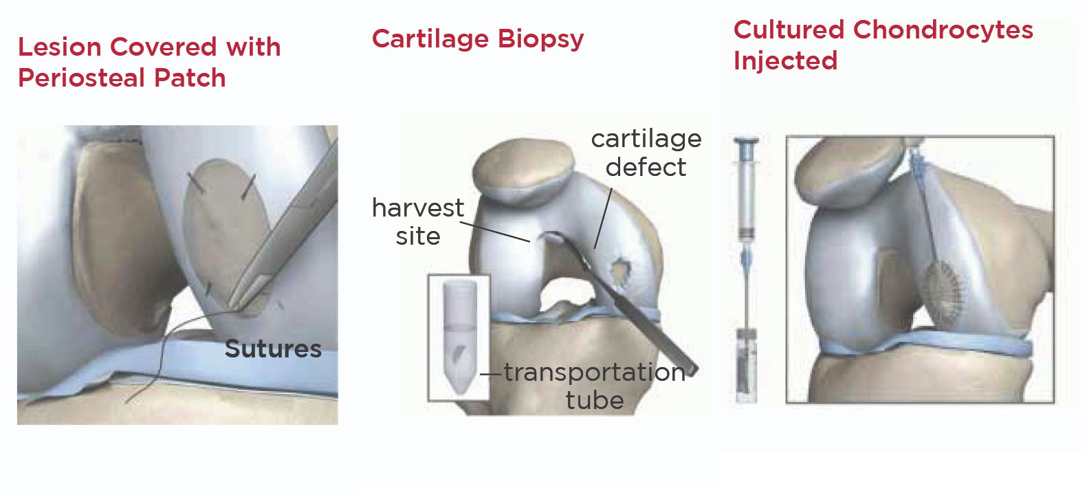
Wouldn’t it be great if we could take a tic-tac size piece of cartilage from your knee, grow your own cartilage in the lab, and restore your joint surface by filling in the defects with your own cells?  Well, at AOS you can!

We can take a small piece of cartilage from your knee joint and grow it in the laboratory.  The grown cells are then attached to a membrane which is then inserted into the defect of the knee, secured with a biologic glue.  Over a several month period of time these cells may grow and fill in the defect, thus biologically restoring the damaged joint surface.  I believe these types of procedures offer the best long-term solution to this kind of knee problem.  I believe that AOS has the most experience and commitment to these type of biological solutions for joint surface defects.

cartilage restoration

If you are between the ages of 15 and 60 and are having problems with pain or swelling in your knee, please ask Dr. Sites or one of the AOS physicians for an evaluation.

May 02, 2017

| AOSHogDocs

Ankle Sprains (Part I of III)

What is an ankle sprain?

An ankle sprain is an injury to the ligamentous structures supporting the ankle.  There are 3 basic types of ankle sprains – lateral, medial and high, with lateral ankle sprains being the most common.

The classic lateral ankle sprain is when the ankle is twisted in such a way the foot turns inward (inversion injury) damaging one of the 3 lateral (outside) ligaments.  The pain and swelling is on the lateral side of the ankle.

A medial ankle sprain occurs when the ankle is twisted in such a way that the foot turns outward (eversion injury) damaging the single medial ligament called the deltoid ligament.  The pain and swelling with this injury if on the medial side of the ankle.

The high (aka syndesmotic) ankle sprain occurs when the ankle is everted and externally rotated usually with a traumatic force placed on the outside of the ankle.  With this injury, pain and swelling are usually diffuse with a moderate amount of swelling in the front of the ankle.  These take the longest to recover from.

What should I do for an ankle sprain?

It all depends on the type of injury sustained and ligaments damaged.  For any type, the RICE principle is always a good place to start.  RICE starts for REST, ICE, COMPRESSION and ELEVATION, but I add an extra R to the mix – REHABILITATION.  I believe active rehabilitation, which includes proprioceptive (balance) training for ankle sprains are key to future function prevention of future injury.

For lateral ankle sprains, a compressive wrap or lace-up ankle brace can be helpful.  However, medial and high ankle sprains may need additional support in the form of a walking boot and potentially crutches.  There will be additional blog posts specifically discussing the different brace/stabilizer options and common rehab principles for ankle sprains.

When should I see a doctor?

Evaluation in the clinic involves ruling out fractures and evaluating stability of the ankle joint itself.  If you are having difficulty bearing weight on that ankle immediately or having significant pain and swelling, then seeing us at Advanced Orthopaedic Specialists can get you back on the road to recovery.

May 12, 2017

| AOSHogDocs

Ankle Sprains: ICE Vs HEAT (Part II of III)

Have a sprained ankle? Now what? Do you apply Ice or Heat to your injury? In the following article we will discuss ice vs heat and the application and benefits of both.

ICE

Ice can be used at any time of the injury process and should be considered especially in the first 72 hours of your ankle sprain. Ice tends to help with inflammation and swelling because it restricts blood flow to the applied area. It is also used a form of pain control because it can numb the area that it is applied to. Ice therapy can consist of several types of applications, regular Ice, Blue Frozen Gel Packs, frozen bag of peas, or ice water bath immersion. One should take precautions to protect the skin and place some sort of a barrier between the ice and the skin and can ice an injury for 15-20 mins every hour as needed for pain and swelling.

HEAT

Heat works in the opposite way and should be considered for injuries that have not occurred in the last 72 hours. Heat applied to an area causes increased blood flow and can ease pain around joints. Heat also helps with pain because it brings warmth to joints and muscles that love to be warm and not cold. Heat can be applied in several forms that may include a heating pad, moist towel, hot tub. One should also take precautions with heat because it can burn sensitive skin and can also lead to increased swelling and possibly delayed healing. Heat can be applied to the ankle for 10-15 mins as needed to help with pain and range of motion.

When should I see a doctor?

If you have been using Ice for longer than 3 days and your ankle sprain has not improved in swelling, tenderness, or pain you should then call and get an appointment to be examined by a Healthcare Professional to rule out a more extensive injury.

As a final note you can always use ice and heat for other injuries, not just an Ankle Sprain. Ice within the first 3 days and heat can be used after. Ice for decreasing swelling and heat for stiff joints and muscles. If you find that these are not working come and see us at Advanced Orthopaedic Specialists for an evaluation of your injury.

May 31, 2017

| AOSHogDocs

Ankle Sprain: Rehabilitation (PART III)

After an ankle sprain, proper rehabilitation is key to returning to activity quickly and to prevent additional injury later. An injury as common as an ankle sprain can have long lasting effects, if not treated properly. Like most athletic injuries, there are 3 keys to proper ankle rehabilitation. Range of motion (ROM), strength and proprioception will be covered in greater detail below.

1. Gaining proper ROM is the first step to recovery from any type of ankle sprain. Both passive and active ROM is necessary and simple exercises such as a towel stretch can help achieve this goal. Using a towel (or a belt) stretched over the ball of the foot, and pulling the ankle into dorsiflexion is an easy exercise to gain passive ROM. An example of an active ROM exercise is the ABCs, writing out the alphabet with the tip of the toe.

2.Improving strength is the next step in the rehabilitation process. Joints have both static and dynamic stabilizers. A ligament, the static stabilizer, takes many weeks to scar down to prevent further injury. The surrounding musculature, the dynamic stabilizer, is key to protecting the ankle when returning to play quickly. The stabilizer muscles of the ankle can support the ankle in addition to external taping and bracing to allow an athlete to begin to return to play before the injury has fully healed, saving valuable weeks in a season or participating in a schedule event they would have otherwise missed. A resistive exercise, such as 4-way ankle exercises with a theraband, is an example of a strengthening exercise.

3. Proprioception is the ability to sense a stimulus from within the body regarding position, motion, and equilibrium. This essentially means the body can tell where a joint is in space and make proper adjustments based on neural feedback. Regarding the ankle, the most common form or proprioceptive rehab involves balance. This can be as simple as standing on one foot and as difficult a standing on an unstable surface while playing catch or other sport specific motions.

These three principles can and do overlap in most rehab exercises but it is important to have a solid foundation in each area before moving to the next. Strengthening an ankle with poor ROM will lead to an altered gait and additional issues further up the kinetic chain. Training for proprioception with insufficient strength can be dangerous and lead to repeat injury. The physical therapist and athletic trainers at Advanced Orthopaedic Specialists are happy to assist you with setting up a home exercise plan or more advanced rehabilitation in our physical therapy clinics in Fayetteville or our brand new Pinnacle Hills location.

March 07, 2019

| AOSHogDocs

2019 Position Statement on Concussions

March is National Brain Injury Awareness Month, and recently the American Medical Society for Sports Medicine (AMSSM) published their latest position statement on concussion in sport.

This paper, updated from 2013, focuses on the current recommendations regarding the evaluation and management of sport related concussions (SRC). The 2019 position statement discusses the current medical literature around the pathophysiology of concussion, the epidemiology, sideline evaluation tools, treatment recommendations, and issues surrounding potential long-term sequela around concussions.

What’s New?

The most recent epidemiologic data estimates 1-1.8 million SRC annually in the US in those between 0-18 year of age, of which 400,000 occur in high school athletes. The evaluation begins before the season, during the pre-participation physical exams where high risk athletes and a detailed concussion history can be identified.

Once a sport-related concussion is suspected and diagnosed, the active rehabilitation of a concussion may begin using a graded exercise protocol. The days of hibernating concussions is gone. There are return-to-learn protocols in addition to return-to-sport protocols.

How to Stay Informed

For additional information, feel free to download the full position statement, learn about our concussion care services, or sign up for our concussion care seminar on March 14, 2019 to learn about the newest information regarding sport-related concussions.