I have a friend that had a total knee replacement and went home the same day. Is it an option for me? | Advanced Orthopaedic Specialists | Fayetteville, AR | Rogers, AR
Advanced Orthopaedic Specialists

February 13, 2018

| Christopher Arnold, MD

I have a friend that had a total knee replacement and went home the same day. Is it an option for me?

Total knee arthroplasties are one of the most common orthopedic procedures done in the United States today. Not only is it a very common procedure, but it also is an extremely rewarding procedure if done appropriately. Studies have shown that facilities and surgeons that perform a high volume of total knees have higher success rates than facilities or surgeons which perform a low volume of total knees. At Advanced Orthopaedic Specialists, we perform greater than 400 total knees a year and these are done at Physicians’ Specialty Hospital. With an increase in volume, Advanced Orthopaedic Specialists have done everything to make the total knee surgery as seamless as possible. There are certain risks associated with total knees, but Advanced Orthopaedic Specialists have a system in place to minimize these risks as best as possible.

Outpatient total knee replacement is becoming very common across the united states, and Advanced Orthopaedic Specialists has embraced this. Not every patient is a candidate for an outpatient total knee; however, in our practice approximately 10% to 15% of the patients are candidates for this. At Advanced Orthopaedic Specialists, we stratify our patients based on risks. There are typically four groups of patients which we look at as outlined below.

1. Healthy patient with no medical comorbidities such as heart disease, diabetes, obesity, who have a great social support at home, have the ability to be independent the day of the surgery, and live in a major city with a major hospital close by.

2. Patients who have minor medical comorbidities who may not be able to be independent the day of the surgery, and may live in a rural area or may not have good family support at home.

3. The patient with significant medical comorbidities, but desires not to go to formal inpatient rehabilitation facility.

4. A patient with multiple medical comorbidities such as obesity, diabetes, heart disease, who does not have family support at home or is not able to obtain the independence within the first three days to go home.

The patients who fit in the first group are potentially candidates for outpatient total knee replacements. These patients are educated about outpatient total knee prior to the procedure. Their procedure is done in the morning and they start physical therapy approximately one hour after the procedure where they are ambulating with assistance. If they are able to obtain independence (dress themselves, get in and out of bed on their own, and ambulate 25 yards), they are considered to be independent.If these patients also are medically stable as manifested by stable vital signs consisting of blood pressure, heart rate, and oxygen saturation, have a stable blood count and exhibit no signs of any blood clot they may be a candidate to go home. Our criteria for outpatient total knee is somebody who is independent, is medically stable, exhibits that they do not have a blood clot as evident on an ultrasound done post-operatively, and has good family support and is not living in a rural area. If patients meet all these criteria, they are candidates to go home. If they are discharged home, they are given a hotline number to call should they experience any issues. They will start physical therapy in an outpatient setting the day after the surgery. They will be discharged home with pain medication, strict instructions on wound care, and instructions on signs and symptoms to watch for, for any medical issues.

In the past year, Advanced Orthopaedic Specialists has performed approximately 40 to 50 outpatient total knees with no complications noted. This is a great aspect of our practice, which allows patients to undergo a major surgery safely and to sleep in their own bed in their own home the night of the surgery.

If you feel you are a candidate for a total knee or an outpatient total knee, please contact Advanced Orthopaedic Specialists.
December 20, 2017

| Christopher Arnold, MD

I Dislocated My Kneecap, What Should I Do?

Patellar dislocations are common injuries that are seen at Advanced Orthopaedic Specialists. A patellar dislocation is whenever the kneecap (patella) dislocates from the femur (trochlea). This is relatively common in teenage athletes. It most commonly occurs with a twisting mechanism with the foot planted in one direction and the body turned in the opposite direction. The athlete will feel their kneecap slide out of the joint. Most commonly, the kneecap will relocate on its own; however, sometimes, it requires a trainer or physician to relocate it.

The patellofemoral joint is a convex-concave articulation. Some patients have less of a concavity on the thigh, which predisposes them to a patellar dislocation. The good news is that the majority of patellar dislocations are treated non-operatively. If a patient is seen at Advanced Orthopaedic Specialists with a patellar dislocation, we initially treat them with a brace, aggressive physical therapy, and anti-inflammatories. Once the swelling and pain has subsided, we will release them to return to sports. This can take anywhere from one to four weeks. The majority of patients who experience patellar dislocation will have no further problems as long as they keep their quadriceps strong and their muscles stretched.

If a patient experiences recurrent dislocation, this prompts us to investigate this further with MRIs. The MRI can show an injury to the main stabilizer of the patellofemoral joint, which is the medial patellofemoral ligament. The more times the patellar dislocates, the more likely there is injury to the medial patellofemoral ligament. This is the main stabilizer of the patellofemoral joint and if the patella continues to dislocate, this would be an indication for surgery. In the past, patellofemoral reconstruction surgeries were very unsuccessful; however, with the advent of reconstruction of the medial patellofemoral ligament, there is approximately a 95% success rate of no further dislocations and return to full activity.

Sometimes, when the patella dislocates, it can injure the cartilage behind the patella or on the adjacent femur. This can sometimes lead to small cracks in the cartilage or even loose cartilage pieces that float within the knee. If this happens the patient would experience pain and swelling and would require an arthroscopy to smooth down the cracks or retrieve any loose pieces.

The good news with the patellar dislocations is that the majority (greater than 90%) are treated with aggressive therapy, anti-inflammatories, and bracing. However, if this fails, the patient can expect a full return with either an arthroscopy to retrieve the loose pieces or a formal reconstruction. If you had episodes of patellar dislocations, call Advanced Orthopaedic Specialists.

September 13, 2019

| Christopher Arnold, MD

I Broke My Collar Bone. What Should I Know?

If you or a family member have broken your collar bone, you may be wondering if it will require surgery and when you can return to normal activities. These are common questions, and we can help address them, as well as outline a few important things you should know.
Your collar bone, also known as the clavicle, is a long, narrow bone, at the top of the shoulder that goes from the sternum (or the chest) to the tip of the shoulder (or the acromion). It is one of the more commonly fractured bones in the upper extremity. Traditionally, these have been treated without surgery. More recently, though, it has been common to treat these surgically with better results.

Where and How Do They Typically Occur?

The most common clavicle fractures occur in the middle portion. They can also occur toward the end of the tip of the shoulder or toward the chest, which is uncommon. These fractures are usually the result of a high-energy mechanism, such as a sport or motor vehicle accident. It is extremely rare to have an associated injury to the nerves or blood vessels in conjunction with a clavicle fracture.

What Are Treatment Options?

In the past, the majority of clavicle fractures were treated nonoperatively with a sling for approximately six weeks. The patient was allowed to resume normal activities once there was good healing, which is typically three to four months.

More recently, there has been a trend toward treating these surgically. Some of the indications for surgical treatment are if the fracture is displaced, significantly separated, or if it is significantly shortened.

A recent study comparing patients with displaced clavicle fractures around the middle portion, receiving either surgical treatment or nonsurgical treatment, showed that the patient in the surgical group had a statistically significantly improved outcome with regard to function with very few complications.

What Are the Pros and Cons of Nonoperative Treatment?

The advantages of nonoperative treatment are that the patient is not exposed to the risk of surgery. such as infection or damage to nerves, blood vessels, and tendons. Although, these are extremely rare complications. The disadvantage of nonoperative treatment is that the fracture may heal in a nonanatomic position, and the patient may experience significant pain during the healing process as it is difficult to completely immobilize the clavicle.

What Are the Pros and Cons of Surgical Treatment?

The advantages of surgical treatment are that the surgeon uses a plate and screws to stabilize the clavicle allowing a more anatomic healing scenario. With the plate and screws, there is immediate stability of the clavicle, which results in improved pain once the pain from the surgery has subsided. The disadvantage of surgery is the risk of infection and/or damage to nerves, blood vessels, and tendons, which we have not seen in our practice.

We’re the Experts in Total Shoulder Care.

At Advanced Orthopedic Specialists, our shoulder surgeons take care of a large number of high-level athletes, as well as recreational athletes. These athletes have typically sought surgical treatment of their clavicle fracture to improve the function of the upper extremity, to diminish the pain, and allow them to return to their desired level of sport in a quicker fashion.

What Should You Expect from Surgery?

This surgery typically takes about one hour. It is done in an outpatient setting. The fracture is reduced anatomically, and the plate is affixed to the fracture to hold it securely. The patient wears a sling until the wound is healed, which is typically 10 to 14 days. Once the wound is healed and the sling is discontinued, physical therapy is started and the patient is allowed to begin running. We begin light strengthening at approximately four to six weeks and allow a return to sports at approximately 7 to 12 weeks, depending on the amount of healing needed. The disadvantage is that some patients do elect to have the plate removed at some point.

If you sustain a clavicle fracture, understand there are options to help you get you back into your sport or normal life activities as soon as possible. Getting you back to your personal best is our ultimate goal.

If you have specific questions or would like one of our physicians to take a look at your collar bone injury, feel free contact us and set up an appointment right away.

January 24, 2017

| AOSHogDocs

Hip Impingement

What is it?

Femoroacetabular impingement (FAI) is a common cause of hip pain. Impingement can occur due to an abnormal shape of the head of the femur (ball) or acetabulum (socket). If there is an abnormal shape of the femur it is called “cam impingement”. In this type of impingement the femur has a bump on it. When the leg is moved in a certain way the bump contacts the socket resulting in hip pain. Cam impingement is more common in young, athletic males. The other type of impingement is called “pincer impingement”. In this type of impingement the acetabulum (socket) of the hip covers too much of the femur. Pincer impingement is more common in middle-aged women. Patients who have impingement are at a higher risk for labral tears in their hip, which is a ring of tissue that surrounds the socket of the hip.

What are the Symptoms?

Patients with impingement may experience groin pain and/or hip pain when they move their leg forward, backward, or when turning side to side. Patients often complain of difficulty sitting. It is not uncommon for patients to experience a clicking or catching sensation.

When Should I See My Doctor?

You should see your doctor if you are having groin or hip pain with activity, especially when bending your hip.

How Can I Prevent Hip Impingement?

There is no way to prevent impingement since it is an anatomical issue. Patients with hip or groin pain should limit or modify activities which cause pain.

When Can I Return to Play?

After diagnosis of impingement athletes are started in exercise programs with the goal of decreasing stress on the hip. Athletes may return to their particular sport once they have good motion, strength, and function of their hip.

November 06, 2017

| AOSHogDocs

Hip Bursitis

What is hip bursitis?

Hip bursitis, also known as greater trochanteric bursitis, is a condition that affects the outside region of the hip. The greater trochanter is the bony prominence on the lateral side of the leg and the attachment for many hip and pelvic muscles. In hip bursitis, that region becomes inflamed and painful.

What causes hip bursitis?

There are many causes, from acute traumatic injuries to overuse etiologies. The most common cause is some form of overuse syndrome. Running, cycling and activities that do not naturally include side-to-side movement can lead to the dysfunction.

What are the classic symptoms of hip bursitis?

Classically, hip bursitis presents as lateral hip pain, not localizing to the groin, which is worse with the inciting activity, direct pressure on greater trochanter (side sleeping), or prolonged sitting. On physical exam, a patient is tender directly over the greater trochanter, and possibly a tight iliotibial (IT) band may be found.

How do you treat hip bursitis?

There are many treatment options for hip bursitis from the conservative to the invasive. A patient can start with oral anti-inflammatories and a home stretching program. If that fails, a round of formal physical therapy with or without a steroid injection can be beneficial. In recalcitrant cases, regenerative medicine injections such as PRP (see previous blog posts) or surgical debridement may be needed.

Should you have any other questions or desire evaluation if you believe you have hip bursitis, please feel free to visit us as Advanced Orthopaedic Specialists.

July 29, 2019

| AOSHogDocs

Heat Illness – Quick Facts

What is it?

Heat illness occurs when the body loses its ability to prevent overheating. Normally the body removes excess heat via four different ways:

1. Evaporation: sweating
2. Convection: heat transferred to the environment by motion such as air or water
3. Conduction: transfer of heat from one object to another via direct contact
4. Radiation: transfer from one object to another without direct contact

Heat illness is a spectrum that ranges from mild (heat cramps) to moderate (heat exhaustion) with the most severe being heat stroke.

What are the Symptoms?

Early signs and symptoms include swelling of the hands and feet, cramps, muscle tightening, muscle spasms, and lightheadedness.

More severe signs and symptoms include confusion, agitation, irritability, lack of coordination, headache, nausea, vomiting, excessive sweating, flushing of skin, and rectal temperature greater than 104º F.

What are the Risk Factors?

• Age: More common in older adults and children
• Exercising in the heat, especially if not acclimated
• Sudden change in environmental temperature
• Heavy clothing and/or sporting equipment
• ADD and ADHD medications: Adderall, Vyvanse, and Concerta
• Common cold or allergy medications with pseudoephedrine and phenylephrine
• Certain medical conditions: uncontrolled diabetes, hypertension, kidney disease, low body weight, and dehydration

What Is the Treatment?

Those with mild symptoms should be treated with fluids and resting in a cool area. Those with more severe signs and symptoms may require whole-body immersion for cooling and transfer to the nearest hospital. Never delay cooling of anyone suspected of having heat illness.

How Can It Be Prevented?

Athletes should acclimate their body to heat and humidity at least 10 days prior to competition. During this process, the body adapts by lowering the core temperature, decreasing resting heart rate, increasing sweat rates, and increasing storage of water within the body.

It is also important to wear light-colored and lightweight clothing while exercising. Activity should also be planned around weather conditions, such as avoiding exercise during the peak temperatures of the day. Everyone should hydrate before, during, and after any activity. The best guide to hydration is thirst.

If you are taking medications and/or supplements, review these with your physician prior to starting any exercise regimen. Also, let coaches know if you’re on a medication that predisposes you to heat illness so they can help keep an eye on the situation.

When Can I Return to Play?

If you have a heat-related illness you should be evaluated by a physician prior to returning to exercise. Once you are cleared to return by your doctor you should begin your exercise in a cool environment with gradually increased activity.

August 05, 2019

| AOSHogDocs

Heat Illness – What You Might Not Know

What is a heat-related injury or illness?

Heat-related injury or illness is a result of activity (work or sport) where a person is exposed to heat to the point that their body is unable to compensate and core body temp rises leading to injury or illness.

Why is it important this time of year?

For fall sports, such as football, cross country or soccer, heat exhaustion most often occurs during the first two weeks of pre-season practice, and the risk rises along with the temperatures during these events. Despite the ongoing rise of awareness campaigns, incidents of heat illness continue to occur every season. According to the CDC, between 1999-2010 there were 8,081 heat-related deaths in the US.

What are the different types of heat-related injury/illness?

The main types of heat illness or injury are:

  • Heat Cramps – The first presentation of heat-related injury with associated muscle cramping due to exercise and heat.


  • Heat Exhaustion – The progression of heat injury to the point that the body shows signs of fatigue, headache, nausea, dizziness or fainting, but the body is still sweating and able to protect itself. 


  • Heat Stroke – This is an emergency and life-threatening situation. At this point, there is a progression from exhaustion to the point the body can no longer compensate and protect itself. Sweating stops, core temp rises >104F (40C), the heart rate increases, and the person starts to become confused, neurogenic compromise begins and can lead to organ failure or death. 


What are common risk factors most people don’t know about?

Many people understand that heavy exercise in the heat or a sudden change in environmental temperature can predispose a person to heat illness. Age is another factor. Heat illness is more common in older adults and children.

Medications for ADD and ADHD, such as Adderall, Vyvanse, and Concerta can make someone more vulnerable to heat exhaustion. The same goes for common cold medications or allergy medications with pseudoephedrine and phenylephrine.

Another risk factor includes certain medical conditions. Uncontrolled diabetes, hypertension, kidney disease, low body weight, and dehydration can all make it easier for someone to suffer from heat illness.

How do I protect myself from heat-related injury/illness?

Heat-related injury/illness is preventable. Proper hydration before, during and after activity is helpful. Limited work/practice during the hottest part of the day, or during heat waves (3 consecutive days with temp >90F). Acclimatization to activity allows the body a gradual get used to the environment, both with work and sports. Using proper loose-fitting attire or limiting athletic or work gear which traps heat and prevents adequate cooling. Frequent breaks to allow core temp to lower, and use of fans or other cooling methods to maintain proper core temp.

For additional questions or to receive a proper physical to ensure you’re ready to take to the field, schedule an appointment with one of our sports medicine physicians today. We’re here to help you perform at your best level.

February 28, 2017

| David Yakin, MD

Hallus Rigidus – “Stiff Big Toe”

Hallux rigidus means “stiff big toe,” which is the main symptom of the disorder. Hallux rigidus is a form of degenerative arthritis, which can cause pain and stiffness in the metatarsophalangeal joint (the joint where your big toe—the hallux—joins your foot).  Because hallux rigidus is a progressive condition (gets worse over time), the toe’s motion decreases as time goes on, making walking or even standing painful. The pain and stiffness may get worse in cold, damp weather, and the joint may become swollen and inflamed. A bump, like a bunion or callus, often develops on the top of the foot and makes wearing shoes difficult.

There is no single cause of hallux rigidus. It may develop because of overuse of the joint, such as in workers who have to stoop and squat or athletes who place a great deal of stress on the joint. It may occur after an injury, such as stubbing the toe or spraining the joint (called “turf toe” in athletes). In some people, hallux rigidus runs in the family and comes from inheriting a foot type or a way of walking that may lead to this condition. Osteoarthritis (wear and tear arthritis) and inflammatory diseases such as rheumatoid arthritis or gout are other possible causes of hallux rigidus.

Your doctor can diagnose the condition by testing the range of motion of the joint—how far the toe can bend up and down. X-rays can show if there are any abnormalities in the bone or bone spur development.

Early treatment of hallux rigidus usually includes the following measures:
• Wearing appropriate shoes with plenty of room for your toes. Some patients find that shoes with very stiff soles relieve pain. Women should avoid wearing high heels.
• Placing pads in your shoe to limit movement of your big toe
• Avoiding high-impact activities, such as jogging
• Taking non-steroidal anti-inflammatory medications, such as ibuprofen, to help relieve the pain and reduce swelling in your big toe. Your doctor may recommend corticosteroid injections into the joint.

If pain and stiffness continue, surgery may be necessary. Shaving the bone spur (cheilectomy) may help relieve the pain and preserve joint motion. Sometimes it may be necessary to cut the bone (osteotomy) in order to realign or shorten the big toe.  If the condition is severe, joint fusion (arthrodesis) may be the best option for long-lasting pain relief.

August 22, 2017

| Zac Snow, DPT

Giving 100% – 10% AT A TIME

When progressing a runner or triathlete back to sport I often emphasize that it is better to undertrain rather than to over-train for a race. The downside to an undertrained endurance athlete is a missed personal record (PR) or feeling winded during their race. On the other hand, the downside to an overtrained endurance athlete is often more debilitating. The result of overtraining in any sport can lead to varying degrees of overuse injuries such as stress fractures, tendinopathies, and other general sprains/strains that can keep the athlete out of their race.
A regimented method of progressing an athlete during training is to use the 10% Rule. This rule states that the athlete’s progression should comprise no more than 10% of their total volume the previous week. For example, while training for a marathon an athlete accumulates 30 miles during Week A. By using the 10% Rule the athlete would increase their mileage by 3 miles to reach a total of 33 miles for Week B.

Now, this 10% volume progression becomes more complicated with multiple sports such as triathlon. It is less likely that the athlete will develop overuse injuries from cycling or swimming when compared to impact sports such as running, but a regimented progression is advised to prevent injury. Increasing 10% of the mileage for running and cycling, and meters/yards for swimming should be done for each discipline. For example, while training for an Olympic distance triathlon the athlete accumulates 1000 meters swimming, 40 miles cycling and 10 miles running during Week C. By using the 10% Rule for each discipline the athlete would accumulate 1100 meters swimming, 44 miles cycling and 11 miles running during Week D.

This progression does not apply to a healthy, experienced endurance athlete. These athletes typically have trained with a coach or independently progressed themselves towards a more rigorous training program that allows them to be more efficient with their time. Athletes new to the endurance realm should utilize the 10% Rule to build a strong base and then become more aggressive with their training or, better yet, get a coach or join a training group.

December 27, 2016

| David Yakin, MD

For the Heel of It! Haglund’s Deformity

What Is Haglund’s Deformity?

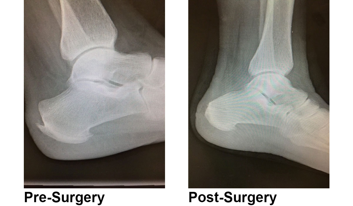
Haglund’s deformity is a bony bump on the back of your heel.  Haglund’s deformity typically develops in people who wear shoes with stiff, closed heels. Your risk of  developing Haglund’s deformity also depends on the shape of your heel bone.
Both surgical and nonsurgical treatments are available to relieve the pain associated with Haglund’s deformity.

Haglund’s deformity is an abnormality of the foot bone and soft tissues. An enlargement of the bony section of your heel (where the Achilles tendon is located) triggers this condition. The soft tissue near the back of the heel can become irritated when the large, bony bump rubs against rigid shoes. This often leads to bursitis. (Bursitis is an inflammation of the fluid-filled sac between the tendon and the bone.) When the heel becomes inflamed, calcium can build up in the heel bone. This makes the bump larger and increases your pain. The condition may worsen causing degenerative tearing of the achilles tendon insertion.

Haglund’s deformity occurs when there’s frequent pressure on the backs of your heels. It may be caused by wearing shoes that are too tight or stiff in the heel. Since it often develops in women who wear pump-style high heels, Haglund’s deformity is sometimes referred to as a “pump bump.”  You may also be more at risk for getting Haglund’s deformity if you have high foot arch, have a tight Achilles tendon, or tend to walk on the outside of your heel.

Haglund’s deformity can occur in one or both feet. The symptoms may include:
-a bony bump on the back of your heel
-severe pain in the area where your Achilles tendon attaches to your heel
-swelling in the bursa, which is the fluid-filled sac at the back of your heel
-redness near the inflamed tissue

Haglund’s deformity can be difficult to diagnose because the symptoms are similar to those associated with other foot issues, including achilles tendonitis.  Your doctor might be able to diagnose the condition based on the appearance of your heel. Your doctor may X-ray of your heel bone if they think you have Haglund’s deformity. This will help your doctor determine whether you have the prominent heel bone associated with the disease.

The treatment for Haglund’s deformity usually focuses on relieving pain and taking pressure off of your heel bone. Nonsurgical options include:
-wearing open-back shoes, such as clogs
-taking non-steroidal anti-inflammatory drugs (NSAIDs), such as ibuprofen or aspirin
-icing the bump for 20 to 40 minutes per day to reduce swelling
-getting ultrasound treatments
-getting a soft tissue massage
-wearing orthotics
-wearing heel pads to reduce pressure from your shoes
-wearing an immobilizing boot or cast

Surgery can also be used to treat Haglund’s deformity if less invasive methods don’t work. During surgery, your doctor will remove the excess bone from your heel. The bone may also be smoothed and filed down. This reduces the pressure on the bursa and soft tissue.

haglunds deformity surgery

You may be given a general anesthesia that will put you to sleep during the surgery. This is usually done if your Achilles tendon is damaged and your doctor needs to fix it.

After surgery, it will take up to 3-4 months for you to get back to all activities including sports. Your doctor will likely give you a boot or cast to protect your foot. You may also need to use crutches for a few days.

The cut will have to remain bandaged for at least seven days. Within two weeks, your stitches will be removed. Your doctor may want to get an X-ray of your foot on follow-up visits to ensure that it’s healing properly.